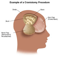
Craniotomy is a procedure in which a surgeon removes a section of the skull and replaces the piece of bone, or bone flap, immediately afterward using titanium screws and plates. In craniectomy, however, they will replace the bone flap much later.
Surgeons perform craniectomy to prevent or relieve severe swelling around the brain, usually due to trauma or a major stroke. They will then replace the bone flap weeks or months later.
Keep reading for more information about how craniotomy is different from craniectomy and burr hole procedures, what to expect from craniotomy, and more.
Craniotomy involves a surgeon removing a piece of the skull to access the brain for brain surgery. They may perform craniotomy for several reasons, including:

- removing a brain tumor
- repairing an aneurysm
- removing a blood clot
- treating epilepsy
- implanting stimulator devices
- repairing skull fractures
- fixing a tear in the membrane lining the brain
A craniectomy is a surgical procedure that is very similar to a craniotomy, but with one key difference. After a craniectomy, the bone fragment is not immediately put back into place. This approach may be taken if there is significant swelling in the brain and a surgeon deems it necessary to relieve pressure within the skull. The bone fragment is typically kept so that it can be put back into place during a future surgery, although it may also be discarded in favor of a future reconstruction using an artificial bone.
A surgeon may perform craniectomy after brain surgery. Craniectomy provides space so that postoperative edema will not cause brain damage due to pressure in the closed skull compartment.
In contrast to craniotomy, the surgeon will not replace the bone right after surgery. Weeks or months later, they will cover the opening with either the original bone flap or a synthetic material during a procedure called cranioplasty.
Craniotomy vs. burr hole
Craniotomy and burr hole are similar procedures. In both cases, a surgeon will remove a part of the skull. Compared with burr hole procedures, craniotomy generally involves removing a larger part of the skull.
According to John Hopkins Medicine, burr holes are smaller openings in the skull for certain surgical procedures, such as the removal of blood buildup due to bleeding.
A surgeon may use a burr hole procedure to treat a subdural hematoma, or bleeding around the brain. This usually occurs due to a fall.
Types
There are several different types of craniotomy. They differ based on location on the skull and the size of the hole the surgeon will make.
Some examples include:
- Supraorbital craniotomy: Often called the eyebrow craniotomy, this procedure involves a surgeon making a hole in the bone just above the eyebrows to access tumors near the front of the head.
- Extended bifrontal craniotomy: During this procedure, a surgeon will remove part of the skull just behind the hairline to access tumors or bleeds at the front of the brain.
- Translabyrinthine craniotomy: During this procedure, a surgeon will remove a portion of the skull in an area behind the ear to access tumors or other lesions in that area.
- Retrosigmoid craniotomy: This is a minimally invasive type of craniotomy wherein a surgeon will make a small hole in the bone behind the ear to remove brain tumors.
- Orbitozygomatic craniotomy: This is a more aggressive type of craniotomy wherein a surgeon will remove a portion of the skull behind the scalp line (near the cheeks) to access deep tumors, such as pituitary tumors.
How to prepare
A person should ask questions about the procedure, especially if they are feeling nervous. They should also tell their doctor if they:
- have any allergies to latex or other surgical materials
- are taking any medications
- have a bleeding disorder
The University of Rochester and Intermountain Healthcare provide some advice on what to expect before craniotomy. Preparation will involve:
- undergoing a physical examination
- undergoing a neurological examination to test against after the procedure
- fasting on the night before the procedure
- undergoing a blood test before the procedure
- taking a sedative to relax the day before the procedure
- shaving the incision area
Procedure
Although practices may vary slightly, the University of Rochester provide some insight into how craniotomy will generally proceed.
An anesthesiologist will monitor the person’s vital signs, and hospital staff will shave the area of the scalp where the surgeon will make the incision.
As the procedure begins, the surgical team will use a device to hold the person’s head in place.
Once the surgeon has made the incisions, they will retract the scalp back to help prevent bleeding. They will then use a drill to create a burr hole or a saw to remove a portion of the skull. They will put this portion of the skull in a sterile solution and cut open the meningeal membrane surrounding the brain.
When the person’s brain is exposed, the surgeon will perform the necessary procedure, such as removing a tumor or an aneurysm. They will then repair the membrane and reattach the bone flap or use a synthetic or metal plate in place of the bone.
Finally, the surgeon will stitch the scalp and dress the wound with a bandage.
What happens immediately after surgery?
Following the procedure, a healthcare professional will take the person to a recovery room. In some cases, they may also provide oxygen.
When the person wakes up, the healthcare professional may transfer them to the intensive care unit (ICU). In the ICU, they will monitor the person’s recovery and provide medication to help ease pain and reduce swelling on the brain.
According to the University of Rochester, a person can expect to stay in the hospital for 3–7 days. In some cases, they may need to go to a rehabilitation center for a few more days.
The hospital should provide the person with detailed instructions on how to care for their incision, as well as what to do at home during their recovery.
Recovery
Once home, a person should follow all instructions from their doctor. These instructions should include how to care for the incision and how to bathe safely.
The incision will likely ache for several days. Therefore, a doctor may suggest taking pain medication — but not aspirin, as this can thin the blood.
The person should slowly return to their usual physical activities, though it will likely take several weeks for them to fully return to the same level of activity as before. They should also avoid lifting heavy objects during recovery. A doctor can recommend when it is best to return to driving.
If the following symptoms occur during the recovery period, the person should contact a doctor:
- any symptoms of infection at the incision site, such as pus, redness, or swelling
- vision changes
- an increase in pain near the incision site
- chills or fever
- confusion
- excessive tiredness
- difficulty speaking
- difficulty breathing
- chest pain
- weakness in the arms or the legs
Risks
Craniotomy is an invasive surgical procedure. For this reason, there are several potential risks, including:
- blood clots
- bleeding
- infection
- seizures
- brain swelling
- muscle weakness
- changes in blood pressure
- pneumonia
- a reaction to the general anesthetic
Craniotomy complications can include:
- speech problems
- paralysis
- coma
A person should talk to their doctor about the risks and complications associated with craniotomy, as they can vary depending on which part of the brain requires the operation.
Does it scar?
The incision the surgeon makes will leave some scarring as it heals, and hair may not grow back in the scar tissue.
This scarring may also contribute to headaches. More than two-thirds of people who have undergone craniotomy experience secondary headaches due to the scarring.
Summary
Craniotomy is a surgical procedure that involves removing a small portion of the skull to provide access to the brain.
A surgeon can then, for example, remove a tumor or release pressure from a bleed.
Following the procedure, a person can expect several days of recovery in the hospital and several weeks of home recovery before returning to their normal activities.
Featured Image Credit: Hopkins Medicine
