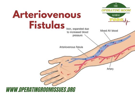
An arteriovenous fistula (AVF) is an abnormal connection between an artery and a vein. When there is a fistula in the brain, it is called an arteriovenous malformation (AVM). When a fistula occurs near the dura (the covering material of the brain), it is a dural arteriovenous fistula. Sometimes AVFs are present at birth (congenital) or develop after birth, and sometimes they are the result of an injury (acquired).
An AVF can occur anywhere in the body, though we mostly find them in the head, neck, spine, and liver. The connection between a high-pressure artery and a low-pressure vein can increase the blood flow through the area, which often expands both the artery and the vein. Often, people with an AVF experience some swelling, pulsing, or vibration in the spot where the AVF is. When a doctor listens to the location with a stethoscope, there is often a bruit, or continuous murmur.
How do I know if I have an AV fistula?
Small AV fistulas that occur in your legs, arms, lungs, kidneys or brain often occur without any symptoms and only need to be monitored by your doctor. Larger AV fistulas may cause:
- Swelling along with a reddish appearance on the skin surface
- Purplish, bulging veins that you can see through your skin, similar to varicose veins
- Swelling in the arms or legs
- Decreased blood pressure
- Fatigue
- Heart failure
An AV fistula in your lung is quite serious and can cause:
- Skin to look blue
- Finger clubbing
- Difficulty breathing (especially when exercising)
- Possible stroke
Causes of AV Fistulas
AV fistulas can develop anywhere in the body, but they typically occur in the legs. They can be caused from:
- Cardiac catheterization complications
- Injuries that pierce the skin
- Genetic conditions
- Congenital defects
Regardless of the cause, serious complications can develop if a large AV fistula is not treated, including heart failure, blood clots, leg pain, and stroke or bleeding.
How are AV fistulas diagnosed?
If you suspect you may have an AV fistula, your doctor will place a stethoscope over the area where a fistulas is suspected and listen for a sound similar to clicking or humming machinery (machinery murmur). If a murmur is heard, he or she will confirm it is caused by AV fistula, using one or more of the following diagnostic tests:
- Duplex ultrasound
- CT ultrasound
- Magnetic resonance angiography (MRA)
Here are some of the most common forms of arteriovenous fistulas:
- Acquired arteriovenous fistulas are not present at birth. They usually happen when a sharp object goes through your body tissue, such as during a gunshot or stabbing injury. These rarely result from a medical procedure.
- Arteriovenous fistulas in the face or neck cause swelling and abnormal pulsing. In extreme cases among infants, it can overload the heart. They rarely cause serious problems with brain development.
- Dural arteriovenous fistulas occur within the dura, which covers the brain. Sometimes we see symptoms at birth while other times, the condition does not become apparent until later in childhood. A large dural AVF can cause cardiac failure at birth. Smaller ones can cause increase in the pressure in the veins inside the head, resulting in hydrocephalus or enlargement of the cerebral ventricles (spaces within the brain that hold fluid). A dural AVF can also cause a bruit, or a pulsing noise or, less often, bleeding or damage to the brain tissue.
- Peripheral arteriovenous fistulas occur outside of the head, neck, and spine. When present at birth, these are most likely in the liver (called hepatic AVF). There are two types of hepatic AVF. A fistula between the hepatic (liver) artery and the hepatic vein is called a hepatic AVF and one between the hepatic artery and the portal vein (the vein that conducts blood from the veins draining the bowel to the liver) is an arterioportal fistula. Both types of AVF can occur within an infantile hemangioma.
Pial or cerebral arteriovenous fistulas occur in the brain. A large one can cause heart failure at or even before birth. Smaller ones can damage the brain around the fistula because it diverts blood flow away from the brain tissue and into the draining vein. We treat these conditions as soon as we discover them, to minimize the amount of brain damage. Some cerebral AVFs rupture, causing bleeding in the brain.- Spinal arteriovenous fistulas happen in or next to the spinal cord, within the spine, or in the muscles adjacent to the spine. These fistulas can compress the spinal cord, leading to numbness or weakness.
- Vein of Galen arteriovenous fistulas usually appear in infancy or early childhood. They drain into the vein of Galen, which is part of the deep venous drainage system of the brain. These fistulas can cause cardiac failure, hydrocephalus, or damage to the developing brain.
Treatment Options
Like arteriovenous malformations, we can treat arteriovenous fistulas with endovascular embolization, microsurgery, or stereotactic radiosurgery. Our multidisciplinary team, which evaluates every complex case, will determine the best approach for you. This team consists of experts in all of our treatment approaches, so can work together to prescribe the safest, most effective treatment plan for your arteriovenous fistula without bias towards one particular treatment.
- Endovascular embolization is the most common form of treatment for an AVF. We perform this procedure by inserting a catheter into an artery (usually the femoral artery in the front of the hip). Then, guided by fluoroscopic or X-ray imaging, we move it to the location of the fistula. We inject contrast so that we can see the exact location of the AVF. Then we inject material into the exact location where the artery and the vein meet, to stop the blood flow. We use a variety of types of devices, including coils, detachable balloons, embolization glue, embolization particles, embolization material (called Onyx), and vascular plugs. Once we have closed the connection between the artery and the vein, the AVF is cured and usually does not reoccur.
Occasionally, when the fistula happens between the side of an important artery and the vein next to it, we insert a covered stent (a wire mesh tube covered with fabric) into either the artery or the vein. This technique typically cures the AVF while keeping the artery and vein intact. A third option is to surgically close the fistula.
- Microsurgery is the most appropriate treatment for a dural, brain, or spinal AVF, either alone or in combination with endovascular embolization. We usually can tell you before we begin treatment if we think microsurgery will be necessary. With microsurgery, we perform a neurosurgery to visualize the AVF under a microscope and we place a titanium clip over the abnormal connection to prevent blood from flowing abnormally from the artery to the vein. During the procedure, we can see the abnormal blood flow stop and the abnormal vein changes from red (when it is carrying arterial blood) to blue, which is the normal color of veins. Immediately after the procedure, we usually repeat the angiogram to confirm that the AVF has been completely treated.
- Stereotactic radiosurgery is appropriate if an AVF is located too close to important brain structures for us to safely perform embolization or microsurgery. This is a painless, outpatient procedure that takes place in the radiation oncology department. We position a stereotactic head frame on you using lidocaine. Music therapy makes the process as comfortable and pain-free as possible. Once the head frame is in place, you undergo a computed tomography (CT) scan of the head. The treatment is very similar to a CT scan and often lasts about 30 minutes. Then we remove the head frame and you can go home.

