Magnetic resonance imaging (MRI) is a type of scan that uses strong magnetic fields and radio waves to produce detailed images of the inside of the body.
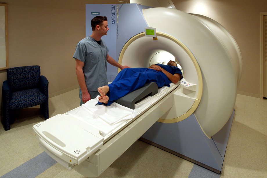
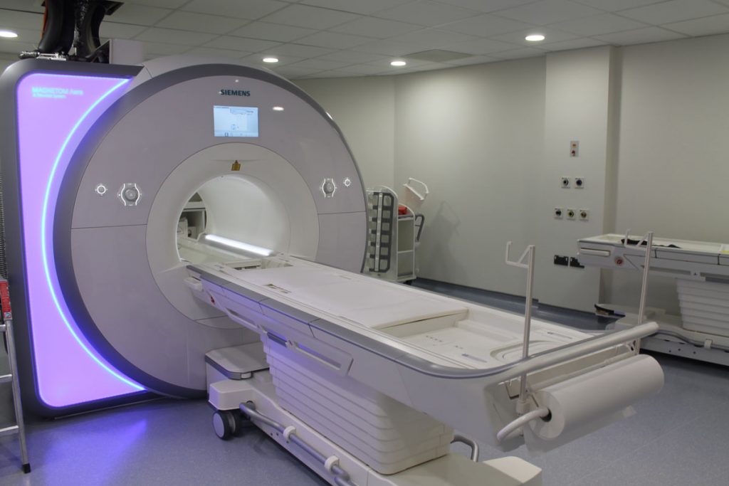
An MRI scanner is a large tube that contains powerful magnets. You lie inside the tube during the scan.
An MRI scan can be used to examine almost any part of the body, including the:
- brain and spinal cord
- bones and joints
- breasts
- heart and blood vessels
- internal organs, such as the liver, womb or prostate gland
The results of an MRI scan can be used to help diagnose conditions, plan treatments and assess how effective previous treatment has been.
What happens during an MRI scan?
During an MRI scan, you lie on a flat bed that’s moved into the scanner.
Depending on the part of your body being scanned, you’ll be moved into the scanner either head first or feet first.

The MRI scanner is operated by a radiographer, who is trained in carrying out imaging investigations.
They control the scanner using a computer, which is in a different room, to keep it away from the magnetic field generated by the scanner.
You’ll be able to talk to the radiographer through an intercom and they’ll be able to see you on a television monitor throughout the scan.
At certain times during the scan, the scanner will make loud tapping noises. This is the electric current in the scanner coils being turned on and off.
You’ll be given earplugs or headphones to wear.
It’s very important to keep as still as possible during your MRI scan.
The scan lasts 15 to 90 minutes, depending on the size of the area being scanned and how many images are taken.
How does an MRI scan work?
Most of the human body is made up of water molecules, which consist of hydrogen and oxygen atoms.
At the centre of each hydrogen atom is an even smaller particle called a proton. Protons are like tiny magnets and are very sensitive to magnetic fields.
When you lie under the powerful scanner magnets, the protons in your body line up in the same direction, in the same way that a magnet can pull the needle of a compass.
Short bursts of radio waves are then sent to certain areas of the body, knocking the protons out of alignment.
When the radio waves are turned off, the protons realign. This sends out radio signals, which are picked up by receivers.
These signals provide information about the exact location of the protons in the body.
They also help to distinguish between the various types of tissue in the body, because the protons in different types of tissue realign at different speeds and produce distinct signals.
In the same way that millions of pixels on a computer screen can create complex pictures, the signals from the millions of protons in the body are combined to create a detailed image of the inside of the body.
Safety
An MRI scan is a painless and safe procedure. You may find it uncomfortable if you have claustrophobia, but most people are able to manage it with support from the radiographer.
Going into the scanner feet first may be easier, although this isn’t always possible.
Extensive research has been carried out into whether the magnetic fields and radio waves used during MRI scans could pose a risk to the human body.
No evidence has been found to suggest there’s a risk, which means MRI scans are one of the safest medical procedures available.
But MRI scans may not be recommended in certain situations. For example, if you have a metal implant fitted, such as a pacemaker or artificial joint, you may not be able to have an MRI scan.
They’re also not usually recommended during pregnancy.
How it’s performed
A magnetic resonance imaging (MRI) scan is a painless procedure that lasts 15 to 90 minutes, depending on the size of the area being scanned and the number of images being taken.
Before the scan
On the day of your MRI scan, you should be able to eat, drink and take any medication as usual, unless you’re advised otherwise.
In some cases, you may be asked not to eat or drink anything for up to 4 hours before the scan, and sometimes you may be asked to drink a fairly large amount of water beforehand. This depends on the area being scanned.
When you arrive at the hospital, you’ll usually be asked to fill in a questionnaire about your health and medical history. This helps the medical staff to ensure you have the scan safely.
Once you have completed the questionnaire, you’ll usually be asked to give your signed consent for the scan to go ahead.
As the MRI scanner produces strong magnetic fields, it’s important to remove any metal objects from your body.
These include:
- watches
- jewellery, such as earrings and necklaces
- piercings, such as ear, nipple and nose rings
- dentures (false teeth)
- hearing aids
- wigs (some wigs contain traces of metal)
Any valuables can usually be stored in a secure locker.
Depending on which part of your body is being scanned, you may need to wear a hospital gown during the procedure.
If you don’t need to wear a gown, you should wear clothes without metal zips, fasteners, buttons, underwire (bras), belts or buckles.
Contrast dye
Some MRI scans involve having an injection of contrast dye. This makes certain tissues and blood vessels show up more clearly and in greater detail.
Sometimes the contrast dye can cause side effects, such as:
- feeling or being sick
- a skin rash
- a headache
- dizziness
These side effects are usually mild and don’t last very long.
It’s also possible for contrast dye to cause tissue and organ damage in people with severe kidney disease.
If you have a history of kidney disease, you may be given a blood test to determine how well your kidneys are functioning and whether it’s safe to proceed with the scan.
You should let the staff know if you have a history of allergic reactions or any blood clotting problems before having the injection.
Anaesthesia and sedatives
An MRI scan is a painless procedure, so anaesthesia (painkilling medication) isn’t usually needed.
If you’re claustrophobic, you can ask for a mild sedative to help you relax. You should ask your GP or consultant well in advance of having the scan.
If you decide to have a sedative during the scan, you’ll need to arrange for a friend or family member to drive you home afterwards, as you won’t be able to drive for 24 hours.
Babies and young children may be given a general anaesthetic before having an MRI scan.
This is because it’s very important to stay still during the scan, which babies and young children are often unable to do when they’re awake.
During the scan
An MRI scanner is a short cylinder that’s open at both ends. You’ll lie on a motorised bed that’s moved inside the scanner.
You’ll enter the scanner either head first or feet first, depending on the part of your body being scanned.
In some cases, a frame may be placed over the body part being scanned, such as the head or chest.
This frame contains receivers that pick up the signals sent out by your body during the scan and it can help to create a better-quality image.
A computer is used to operate the MRI scanner, which is located in a different room to keep it away from the magnetic field generated by the scanner.
The radiographer operates the computer, so they’ll also be in a separate room to you.
But you’ll be able to talk to them, usually through an intercom, and they’ll be able to see you at all times on a television monitor.
A friend or family member may be allowed to stay with you while you’re having your scan. Children can usually have a parent with them.
Anyone who stays with you will be asked if they have a pacemaker or any other metal objects in their body.
They’ll also have to follow the same guidelines regarding clothing and the removal of metallic objects.
To avoid the images being blurred, it’s very important to keep the part of your body being scanned still throughout the whole of the scan until the radiographer tells you to relax.
A single scan may take from a few seconds to 3 or 4 minutes. You may be asked to hold your breath during short scans.
Depending on the size of the area being scanned and how many images are taken, the whole procedure will take 15 to 90 minutes.
The MRI scanner will make loud tapping noises at certain times during the procedure. This is the electric current in the scanner coils being turned on and off. You’ll be given earplugs or headphones to wear.
You’re usually able to listen to music through headphones during the scan if you want to, and in some cases you can bring your own CD.
You’ll be moved out of the scanner when your scan is over.
After the scan
An MRI scan is usually carried out as an outpatient procedure. This means you won’t need to stay in hospital overnight.
After the scan, you can resume normal activities immediately. But if you have had a sedative, a friend or relative will need to take you home and stay with you for the first 24 hours.
It’s not safe to drive, operate heavy machinery or drink alcohol for 24 hours after having a sedative.
Your MRI scan needs to be studied by a radiologist (a doctor trained in interpreting scans and X-rays) and possibly discussed with other specialists.
This means it’s unlikely you’ll get the results of your scan immediately.
The radiologist will send a report to the doctor who arranged the scan, who will discuss the results with you.
It usually takes a week or two for the results of an MRI scan to come through, unless they’re needed urgently.
Who can have one?
Magnetic resonance imaging (MRI) is very safe and most people are able to have the procedure.
But in some instances an MRI scan may not be recommended.
Before having an MRI scan, you should tell medical staff if:
- you think you have any metal in your body
- you’re pregnant or breastfeeding
The strong magnets used during the scan can affect any metal implants or fragments in your body.
MRI scans aren’t usually recommended for pregnant women.
Although they’re thought to be generally safe to use in later pregnancy (after 3 months), it’s not known whether the strong magnetic fields have any long-term effects on the developing baby.
Metal implants or fragments
Having something metallic in your body doesn’t necessarily mean you can’t have an MRI scan, but it’s important for medical staff carrying out the scan to be aware of it.
They can decide on a case-by-case basis if there are any risks, or if further measures need to be taken to ensure the scan is as safe as possible.
For example, it may be possible to make a pacemaker or defibrillator MRI-safe, or to monitor your heart rhythm during the procedure.
You may need to have an X-ray if you’re unsure about any metal fragments in your body.
Examples of metal implants or fragments include:
- a pacemaker – a small electrical device used to control an irregular heartbeat
- an implantable cardioverter-defibrillator (ICD) – a similar device to a pacemaker that uses electrical shocks to regulate heartbeats
- metal plates, wires, screws or rods – used during surgery for bone fractures
- a nerve stimulator – an electrical implant used to treat long-term nerve pain
- a cochlear implant – a device similar to a hearing aid that’s surgically implanted inside the ear
- a drug pump implant – used to treat long-term pain by delivering painkilling medication directly to an area of the body, such as the lower back
- brain aneurysm clips – small metal clips used to seal blood vessels in the brain that would otherwise be at risk of rupturing (bursting)
- metallic fragments in or near your eyes or blood vessels (common in people who do welding or metalwork for a living)
- prosthetic (artificial) metal heart valves
- penile implants – used to treat erectile dysfunction (impotence)
- eye implants – such as small metal clips used to hold the retina in place
- an intrauterine device (IUD) – a contraceptive device made of plastic and copper that fits inside the womb
- artificial joints – such as those used for a hip replacement or knee replacement
- dental fillings and bridges
- tubal ligation clips – used in female sterilisation
- surgical clips or staples – used to close wounds after an operation
Tattoos
Some tattoo ink contains traces of metal, but most tattoos are safe in an MRI scanner.
Tell the radiographer immediately if you feel any discomfort or heat in your tattoo during the scan.
